LECTURES IN ORAL & MAXILLOFACIAL RADIOLOGY - Principles and Practice for Dental Students and Practitioners,
(2 videos / 16 lectures)

Dr. Frederiksen earned his DDS degree from University of Minnesota and Ph.D (Radiation Biology) from SUNY at Buffalo, Buffalo, NY. He is formerly Professor and Director of Oral and Maxillofacial Radiology at Baylor College of Dentistry, TAMUS-HSC and founder of its Maxillofacial Imaging Center. He is a Diplomate and past President of the American Board of Oral and Maxillofacial Radiology.

Dr. Hui Liang earned her DDS degree and Ph.D (Prosthodontics) from Peking University School of Stomatology, P.R. China and her Master’s degree (Oral and Maxillofacial Radiology) from the University of North Carolina at Chapel Hill, NC. She is currently tenured professor in the Department of Diagnostic Sciences, Texas A&M University College of Dentistry. She is a Diplomate of the American Board of Oral and Maxillofacial Radiology and past Treasurer of the American Academy of Oral and Maxillofacial Radiology.

Lecture 1: Nature of Radiation. The atom has long been considered to be the basic unit of matter. Video 1.
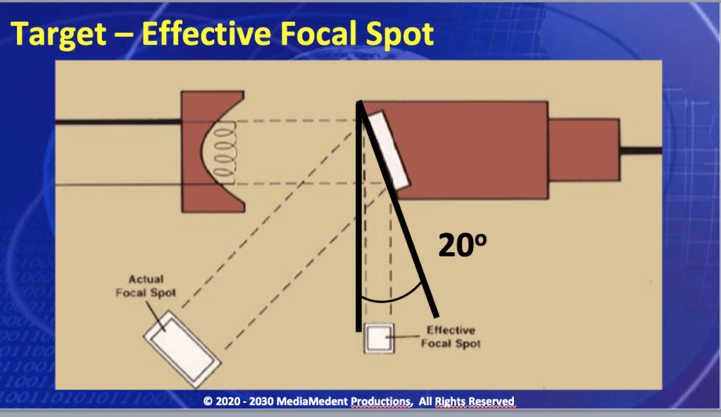
Lecture 2: Production of X Rays. The geometry of the target is one factor in ultimately determining radiographic image quality. Video 1.
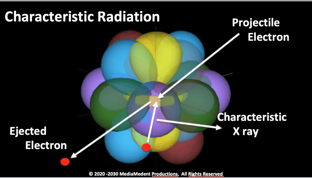
Lecture 3: Characteristics of the X Ray Beam. The second way in which X rays are generated in an X-ray tube is through an ionization interaction that produces characteristic radiation. Video 1.
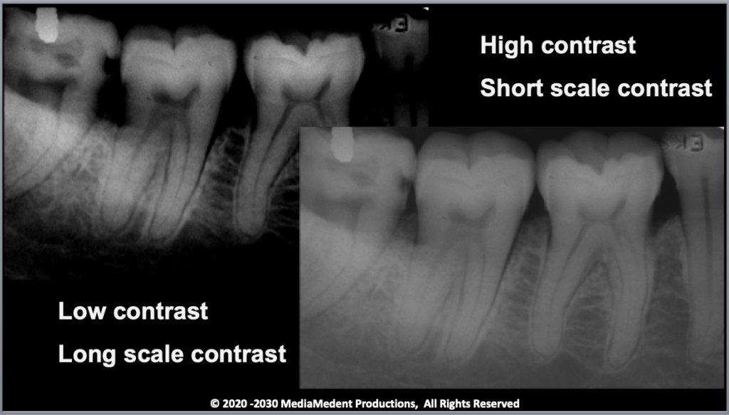
Lecture 4: Photographic and Geometric Properties of the Image. The level of image contrast for most clinicians is a personal preference no matter if the images are recorded using X-ray film or a digital format. Video 1.
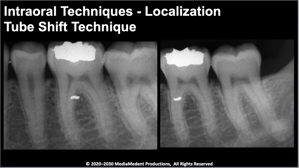
Lecture 5: Techniques in Oral and Maxillofacial Radiology. Relative to the known location of teeth, lingually placed objects, those closer to the tongue, are further away from the X-ray tube. Video 1.
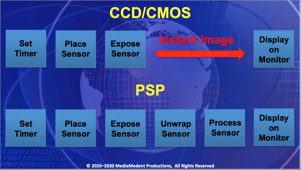
Lecture 6: Digital Radiography. This illustrates the steps involved with obtaining an image using CCD/CMOS and PSP image receptors. Video 1.
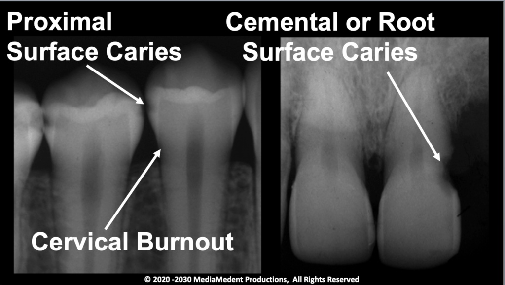
Lecture 7: Teeth and Their Supporting Structures. You should expect to see cervical burnout associated with the cervical region of almost all teeth that have a good level of bone support. Video 1.
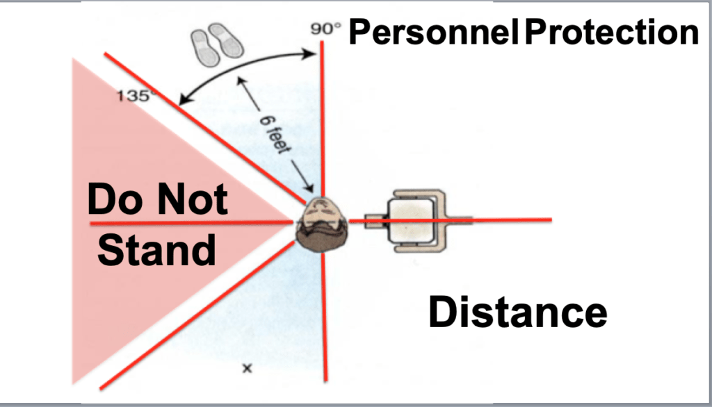
Lecture 8: Radiation Safety. When the exposure is made without any barrier you should be standing at least six feet from the patient. Video 1.
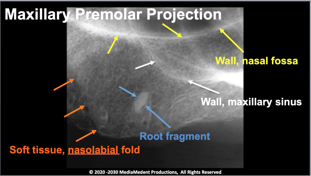
Lecture 9: Landmarks in Intraoral and Panoramic Images. This is a self-paced study. Each illustration will begin with a radiographic image of the jaw. After a pause you will see the illustration labeled with an anatomic landmark. After another pause the landmark will be located. Pause the video as long as necessary to become familiar with the location and appearance of the landmarks. There is no audio associated with this lecture. Video 2.
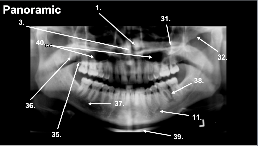
Lecture 10: Landmarks in Intraoral and Panoramic Images – Quiz. This lecture is intended to be a self-paced quiz of anatomic landmarks found in intraoral and panoramic images. Pause the video at any time to study the image. There is no audio associated with this video. Video 2.
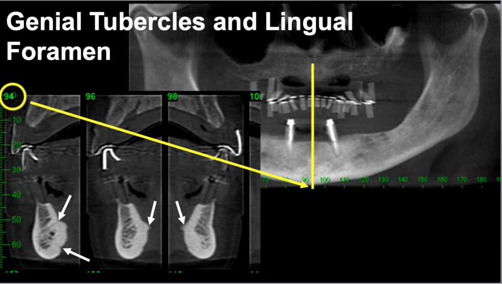
Lecture 11: Landmarks in Cone Beam Computed Tomography and Cephalometric Radiography. Within the image of the genial tubercles can be seen a radiolucency representing the lingual foramen. An accessory foramen may be seen closer to the inferior border of the mandible. Theri is no audio associated with this lecture. Video 2.
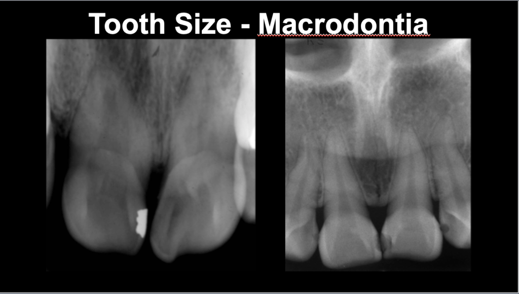
Lecture 12: Developmental Anomalies – Part 1 – Eruption, Number, and Size of Teeth. In macrodontia, illustrated in the image to your left, the teeth appear larger than normal. By comparison, teeth of normal size are as shown in the illustration to your right. Video 2.
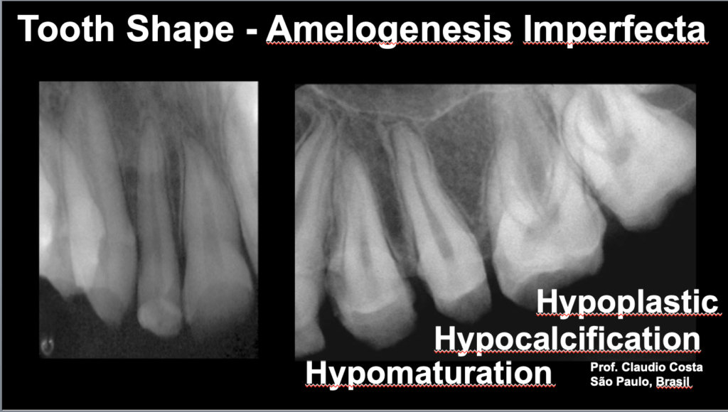
Lecture 13: Developmental Anomalies – Part II – Shape of Teeth. Amelogenesis imperfect is a genetic disorder that interferes with the formation of enamel. Because the disorder is genetic it affects all or nearly all teeth of both the primary and permanent dentitions. Video 2.
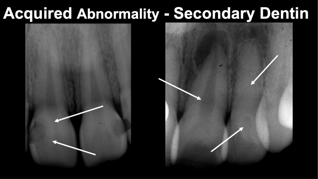
Lecture 14: Dental Anomalies – Acquired. Secondary dentin develops in aging and in response to trauma, attrition, abrasion, and other stimuli. Video 2
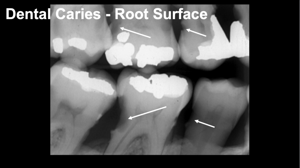
Lecture 15: Dental Caries and Periodontal Disease. When there is gingival recession accompanied by loss of the tooth’s periodontal attachment and loss of alveolar bone, the tooth’s root is vulnerable to root surface or cementum caries. Video 2.
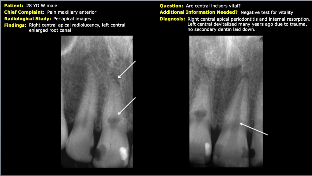
Lecture 16: Comprehensive Self Study Review. A 28 YO W male presented with pain located in the maxillary anterior. What are your findings and radiographic diagnosis? There is no audio associated with this lecture. Video 2.