LECTURES IN ORAL RADIOLOGY - Principles and Practice for Dental Assistants (2 videos / 8 lectures)

Dr. Frederiksen earned his DDS degree from University of Minnesota and Ph.D (Radiation Biology) from SUNY at Buffalo, Buffalo, NY. He is formerly Professor and Director of Oral and Maxillofacial Radiology at Baylor College of Dentistry, TAMUS-HSC and founder of its Maxillofacial Imaging Center. He is a Diplomate and past President of the American Board of Oral and Maxillofacial Radiology.

Dr. Hui Liang earned her DDS degree and Ph.D (Prosthodontics) from Peking University School of Stomatology, P.R. China and her Master’s degree (Oral and Maxillofacial Radiology) from the University of North Carolina at Chapel Hill, NC. She is currently tenured professor in the Department of Diagnostic Sciences, Texas A&M University College of Dentistry. She is a Diplomate of the American Board of Oral and Maxillofacial Radiology and past Treasurer of the American Academy of Oral and Maxillofacial Radiology.
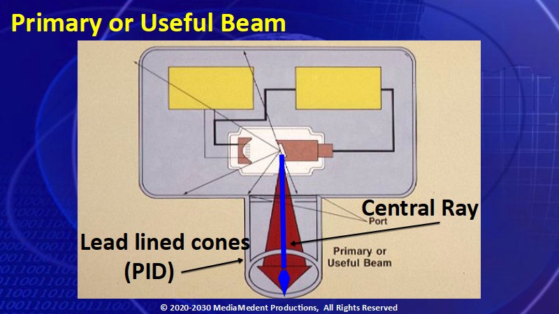
Lecture 1: The Generation of X Rays. X-Rays merge from the X-ray tube as a constantly expanding cone-shaped beam of X-Rays. Because of this characteristic the diameter or size of the primary beam must be restricted by lead lined cones that are called position indicating devices or PIDs. The central ray consists of those X Rays at the geometric center of the beam. Video 1
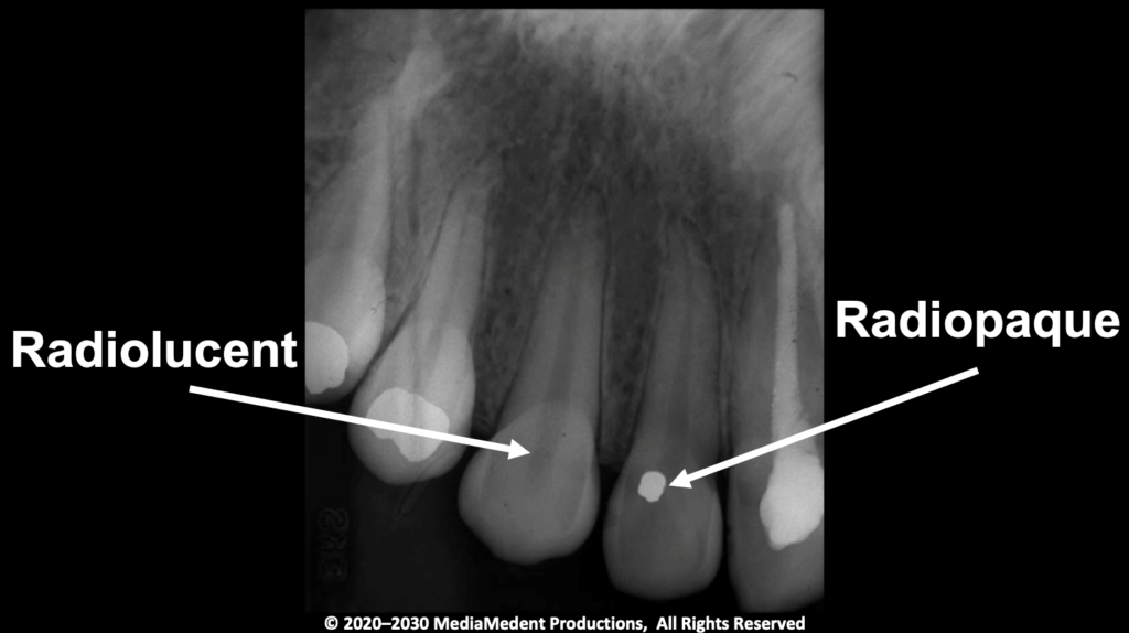
Lecture 2: The Radiographic Image. Radiolucent is a term given to the relatively dark areas in a radiographic image while radiopaque is the term given to the relatively light areas in a radiographic image. Video 1.
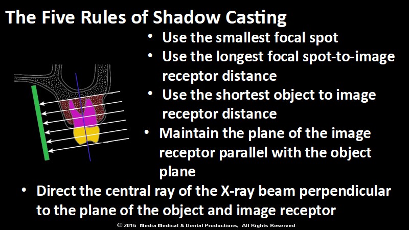
Lecture 3: Techniques in Intraoral and Extraoral Imaging. To obtain the image of an object that most accurately represents the actual size and shape of the object requires an understanding of the five rules of shadow casting. Video 1
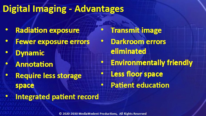
Lecture 4: Digital Radiography. Digital imaging has many advantages over film radiography. Video 1
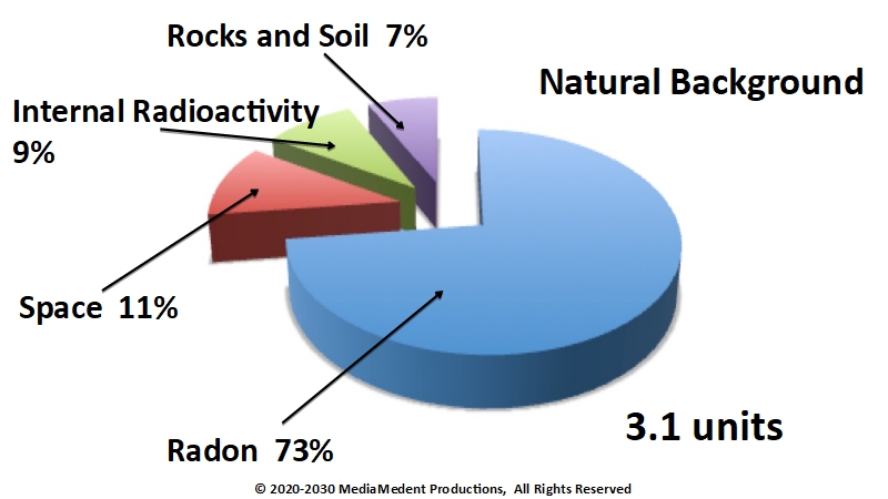
Lecture 5: Radiation Safety. Most natural background radiation comes from radon, but there are other contributions from space, ingested radioactivity, and the rocks, soil, and building materials of our surroundings. Video 1
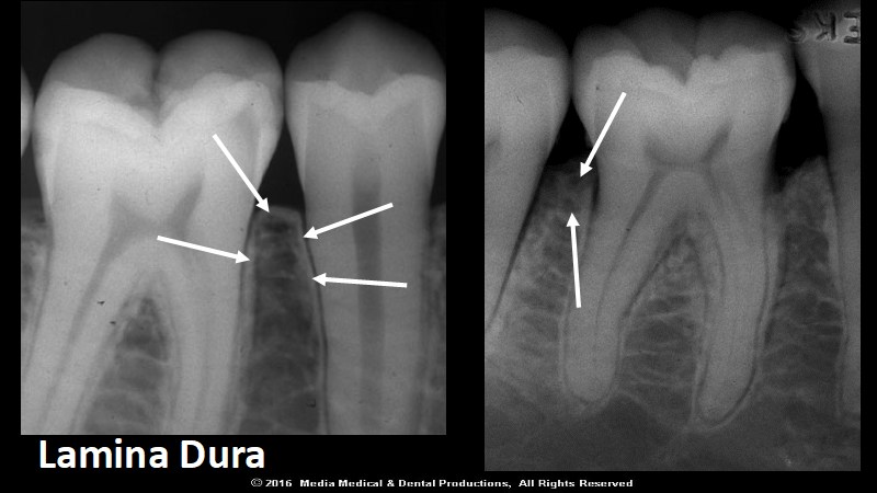
Lecture 6: Teeth and Their Supporting Structures. In the illustration to your left, the lamina dura proceeds superiorly over the alveolar bony crest and then descends alongside the adjacent tooth. Loss of the normal radiographic appearance of the alveolar crest, as shown in the illustration to your right may indicate the onset of periodontal disease. Video 2
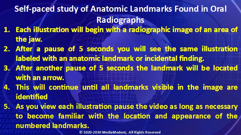
Lecture 7: Landmarks in Intraoral and Panoramic Images. This lecture is intended to be a self-paced study of anatomic landmarks found in oral radiographs. There is no audio associated with this lecture. Video 2

Lecture 8: Landmarks in Intraoral and Panoramic Images. This lecture is intended to be a self-scored quiz of information presented in Lecture 7. There is no audio associated with this lecture. Video 2
